Cartilage Health & Preservation: How to Protect Your Joints

Quick answer
Articular cartilage is the smooth, slippery tissue covering bone ends inside joints; it has no blood supply of its own, so once significantly damaged it has very limited ability to heal. The best protection is regular movement, strong muscles around the joint, a healthy body weight, and prompt attention to joint injuries. Where cartilage is already damaged, treatment ranges from load management and physiotherapy to surgical cartilage-restoration procedures in selected younger patients — assessed at VinayakM in Greater Kailash-1 by an orthopaedic surgeon.
Last reviewed:
July 5, 2026
A cyclist on a morning ride, illustrating low-impact exercise that keeps joint cartilage healthy.

Overview

Articular cartilage is the pearly-white tissue, a few millimetres thick, that caps the ends of bones inside joints such as the knee, hip and ankle. It does two remarkable things at once: it makes joint surfaces more slippery than ice on ice, and it spreads load so that the bone underneath is not damaged by everyday impact.

Cartilage has an unusual biology. It contains no blood vessels and no nerves, and its cells (chondrocytes) are sparse and slow to divide. It is nourished mainly by joint fluid, which is pumped in and out as the joint moves — one reason movement is genuinely food for cartilage. The same biology explains its weakness: with no blood supply, cartilage cannot mount the healing response other tissues use, so significant damage tends to be permanent and can progress towards osteoarthritis over the years.

Understanding this one fact — cartilage protects you, but very little protects cartilage — is the basis of joint care at every age.

Cross-section diagram of a joint showing smooth articular cartilage, a focal cartilage defect and generalised cartilage wear.

Symptoms & signs

Cartilage problems are silent at first because cartilage itself has no nerve endings. Signs appear when damage affects the surrounding joint:

  • Aching after activity, especially loading activities such as stairs, squatting or long walks.
  • Swelling that appears with use and settles with rest.
  • Catching, grinding or clicking as the joint moves over a roughened surface.
  • A localised area of pain in the joint, particularly after a past injury.
  • In more advanced wear, the pattern merges into that of osteoarthritis — stiffness, persistent pain and reduced movement (see our knee osteoarthritis page).

Because early damage is often silent, a history of joint injury — a twisted knee, a kneecap dislocation, a fracture into a joint — is itself a reason to look after that joint deliberately.

Causes & risk factors

Cartilage is damaged in two broad ways:

Sudden injury (focal damage):

  • A direct blow or twist — sports injuries, falls and road accidents can shear off a piece of cartilage, sometimes with underlying bone (an osteochondral injury).
  • Kneecap dislocation frequently damages cartilage on the kneecap or its groove.
  • Fractures that extend into a joint disrupt the smooth surface.
  • Injuries to stabilising structures — an ACL or meniscus tear — change joint mechanics and accelerate cartilage wear over the following years even after the original injury settles.

Gradual overload (wear-related damage):

  • Excess body weight — the strongest modifiable factor; load multiplies across millions of steps.
  • Malalignment — bow legs or knock knees concentrate force on one compartment.
  • Repetitive heavy loading — occupations with constant squatting, kneeling or heavy lifting.
  • Muscle weakness — weak muscles absorb less shock, passing it to the joint surface.
  • Age and genetics — cartilage stiffens and its repair capacity declines with age; family history influences cartilage quality.

Inactivity is also a cause: unloaded cartilage thins, because the fluid pumping that nourishes it stops.

When to see a doctor

See a doctor about a joint if:

  • Pain or swelling follows a significant injury — especially a twist with rapid swelling, a pop, or a kneecap that visibly moved out of place.
  • The joint catches, locks or gives way — a loose cartilage fragment may be present.
  • There is repeated swelling after activity in the same joint.
  • Aching in one joint is persistent or progressive over weeks despite sensible self-care.
  • You had a joint injury in the past and that joint is now becoming gradually more painful — early assessment gives more options.
  • The joint is hot, red and swollen or you feel feverish — this needs urgent care to rule out infection.

Cartilage damage caught early, particularly in younger people, can sometimes be treated before it enlarges — late damage mostly cannot.

How it's diagnosed

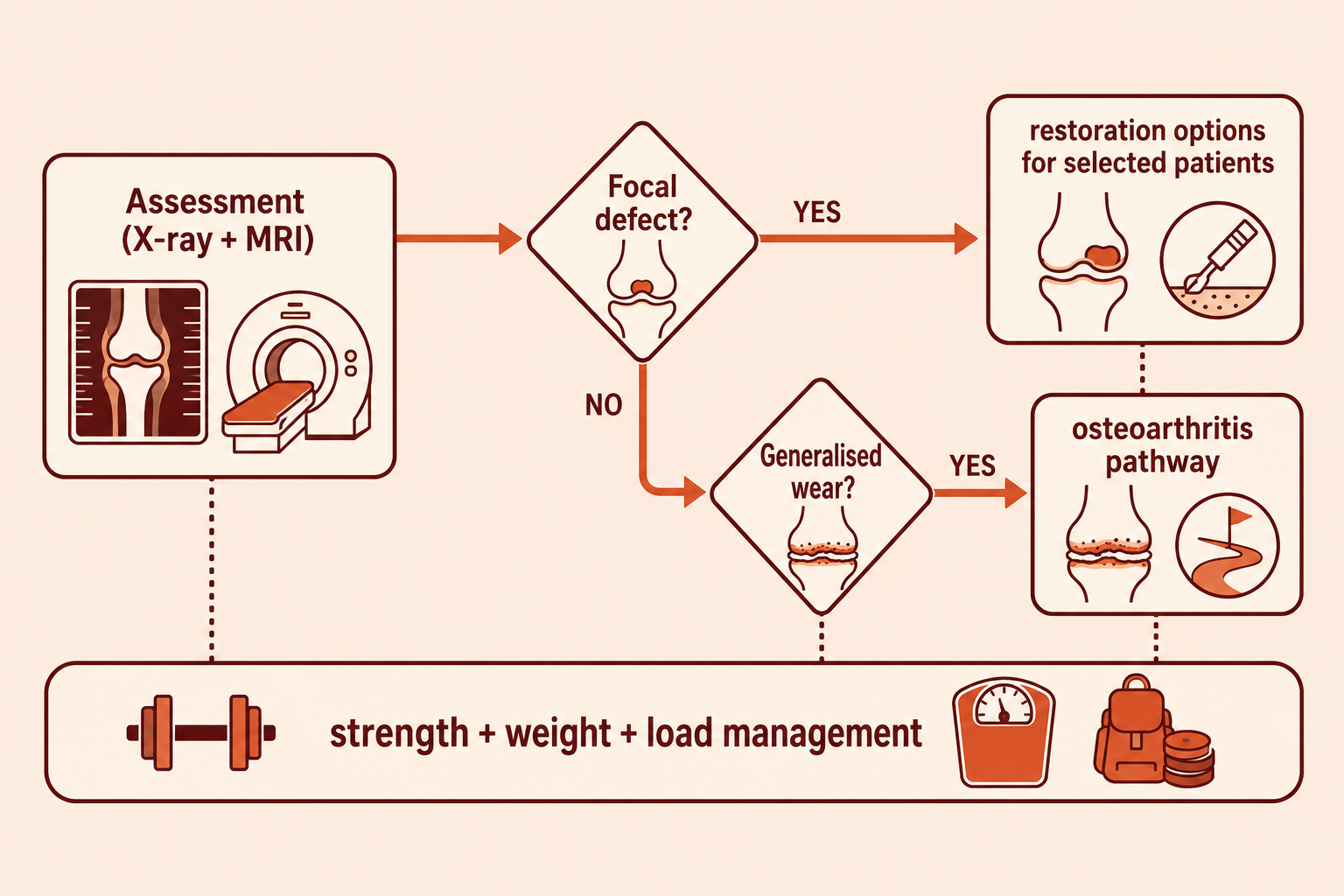
At VinayakM, assessment of suspected cartilage damage includes:

  1. History — past injuries and dislocations, the pattern of pain and swelling, your activity level and goals.
  2. Examination — swelling, localised joint-line or kneecap tenderness, alignment of the limb, muscle strength, stability, and how you walk and squat.
  3. Standing X-rays — to assess joint-space narrowing, alignment and bone changes. Early cartilage damage does not show on X-ray, so a normal X-ray does not rule it out.
  4. MRI — the main tool for seeing cartilage itself: the location, size and depth of damage, the state of the underlying bone, and associated meniscus or ligament injuries.

The assessment establishes whether the problem is a focal defect (a pothole in otherwise good cartilage — potentially repairable) or generalised wear (early osteoarthritis — managed differently), because the treatments are quite different.

Treatment options

There is no medicine that regrows cartilage. Honest treatment is about protecting what remains, and, in selected cases, surgically resurfacing a focal defect.

1. Protect and strengthen (everyone):

  • Load management — temporarily reduce the specific activities that flare the joint, then rebuild gradually.
  • Physiotherapy and strengthening — strong muscles are the joint's shock absorbers; this is the foundation of every cartilage plan.
  • Weight reduction where relevant — the most powerful lever for slowing wear.
  • Low-impact fitness — cycling, swimming and walking maintain cartilage nutrition without overloading it.

2. Symptom control:

  • Topical or short-course oral anti-inflammatories for flares, on medical advice.
  • Supplements such as glucosamine and chondroitin are widely sold; the overall evidence for meaningful benefit is weak, and guidelines such as NICE do not recommend them — we would rather you spend the effort on exercise and weight.

3. Surgical options for focal defects (selected, usually younger patients):

  • Arthroscopic debridement — smoothing loose flaps that catch.
  • Marrow-stimulation (microfracture) — small perforations to encourage repair tissue; the repair is fibrocartilage, less durable than the original.
  • Osteochondral grafting (OATS/mosaicplasty) — moving small plugs of healthy cartilage and bone into the defect.
  • Cell-based procedures (such as ACI) — cartilage cells grown and re-implanted, available in selected centres for suitable defects.

Each option has specific indications, rehabilitation demands and honest limits — none recreates perfect native cartilage, and the right choice depends on the defect, your age, alignment and goals.

4. When wear is generalised, treatment follows the osteoarthritis pathway — see knee osteoarthritis.

How VinayakM helps

At VinayakM in Greater Kailash-1, cartilage problems are assessed by Dr Udit Vinayak, trauma, sports medicine and joint replacement surgeon. That breadth matters for cartilage in particular, because the right answer spans the whole spectrum — from a physiotherapy-and-weight plan, through keyhole procedures for focal defects, to realistic advice when the damage is beyond repair. In practice:

  • Careful distinction between a repairable focal defect and generalised wear, using examination, standing X-rays and MRI where indicated.
  • An exercise-first protection plan for every patient, with dietician support for weight management when useful.
  • For suitable focal defects, a clear discussion of surgical cartilage procedures — what each can and cannot achieve, and the rehabilitation involved.
  • No promises of regrown cartilage — and a warning against clinics that make them.

The goal is decades more service from the joint you have.

Decision pathway for cartilage problems: distinguish focal defect from generalised wear, protect and strengthen, and surgical restoration for selected defects.

Prevention & self-care

Protecting cartilage is mostly about habits sustained over years:

  • Move daily. Cartilage is nourished by movement — regular low-impact activity such as walking, cycling and swimming keeps it healthy. Prolonged inactivity thins it.
  • Keep muscles strong, especially the thighs and hips for the knee. Strength work twice a week is a realistic target.
  • Hold a healthy weight — the single strongest protective factor for weight-bearing joints.
  • Respect joint injuries. After a significant twist, dislocation or fracture, complete the full rehabilitation — an unstable or weak joint quietly grinds its cartilage for years.
  • Increase training gradually — sudden spikes in running distance or new sports are when overload damage happens.
  • Vary your loading. Mix impact and non-impact activity rather than repeating identical heavy loading daily.
  • Don't chase supplements. No pill has been shown to rebuild cartilage; strength, weight and sensible loading do the real work.
Illustration of cartilage-friendly habits: cycling, strength training and maintaining a healthy weight.

Frequently asked questions

Can cartilage grow back on its own?

Essentially no. Articular cartilage has no blood supply, so significant damage does not heal the way skin or bone does. Small areas can fill with a scar-like fibrocartilage, which is less durable than the original. This is why protecting cartilage — through strength, weight control and sensible loading — matters so much.

Do glucosamine or collagen supplements rebuild cartilage?

No supplement has been convincingly shown to rebuild cartilage. Trials of glucosamine and chondroitin show at best small, inconsistent effects on symptoms, and guidelines such as NICE do not recommend them. Exercise, muscle strength and weight management have far stronger evidence.

Is running bad for my cartilage?

For most people with healthy joints, no — recreational running is not shown to wear out knees, and regular loading actually keeps cartilage nourished. Problems arise with sudden increases in distance, running through an injured or malaligned joint, or very high training volumes. Build up gradually and address pain early.

What is a cartilage defect in the knee?

It is a localised area — like a pothole — where the smooth joint-surface cartilage has been damaged, often after an injury or kneecap dislocation, while the rest of the surface remains healthy. Focal defects behave differently from generalised wear and, in selected younger patients, can be treated with cartilage-restoration surgery.

How do I know if I have cartilage damage?

Suspicion comes from the story: aching and swelling after activity, catching or clicking, or a past joint injury. Examination localises the problem, X-rays assess overall wear and alignment, and MRI is the test that actually shows cartilage. A normal X-ray does not exclude early cartilage damage.

Can cartilage damage be cured with an injection?

No injection regrows cartilage. Some injections may ease symptoms in selected situations, but claims that an injection will restore your cartilage are not supported by evidence. Be cautious of any clinic promising cartilage regrowth.

Related reading

References

  1. American Academy of Orthopaedic Surgeons — OrthoInfo. Articular cartilage restoration. — https://orthoinfo.aaos.org/en/treatment/articular-cartilage-restoration/
  2. National Institute for Health and Care Excellence (NICE). Osteoarthritis in over 16s: diagnosis and management. NICE guideline NG226. — https://www.nice.org.uk/guidance/ng226
  3. Towheed T, et al. Glucosamine therapy for treating osteoarthritis. Cochrane Database of Systematic Reviews. 2005;(2):CD002946. — https://doi.org/10.1002/14651858.CD002946.pub2
  4. Fransen M, et al. Exercise for osteoarthritis of the knee. Cochrane Database of Systematic Reviews. 2015;(1):CD004376. — https://doi.org/10.1002/14651858.CD004376.pub3
This page is for general information and education only. It is not a substitute for a consultation, diagnosis or treatment from a qualified clinician. If you have any of the red-flag symptoms above, seek medical care promptly.
{"@context":"https://schema.org","@graph":[{"@type":["MedicalClinic","LocalBusiness"],"@id":"https://www.vinayakm.in/#clinic","name":"VinayakM","url":"https://www.vinayakm.in","logo":"https://cdn.prod.website-files.com/6908cd2bb6a88638d8c0e611/6a494a210e7a3f8e0c33192a_vinayakm-name-logo.png","telephone":"+91 92171 75397","email":"info@vinayakm.in","address":{"@type":"PostalAddress","streetAddress":"3rd floor, B-23, Block B, N Block Market","addressLocality":"Greater Kailash-1, New Delhi","addressRegion":"Delhi","postalCode":"110048","addressCountry":"IN"},"geo":{"@type":"GeoCoordinates","latitude":28.556354,"longitude":77.2323718},"openingHours":["Mo-Tu 09:00-20:00","Th-Su 09:00-20:00"],"founder":{"@type":"Person","name":"Mani Sharma"},"sameAs":["https://maps.app.goo.gl/jPVPXKfH8qAUUDeo8"]},{"@type":"MedicalWebPage","@id":"https://www.vinayakm.in/orthopaedics/cartilage-health#webpage","url":"https://www.vinayakm.in/orthopaedics/cartilage-health","name":"Cartilage Health: Protect Your Joints | VinayakM","description":"Cartilage cushions your joints but has little ability to heal. Learn what damages it, how to protect it, and the treatment options for cartilage damage.","inLanguage":"en-IN","about":{"@type":"MedicalCondition","name":"Articular cartilage damage","alternateName":["Cartilage wear","Chondral damage","Cartilage injury"]},"author":{"@type":"Physician","name":"Dr Udit Vinayak","jobTitle":"Trauma, Sports Medicine & Joint Replacement Surgeon","url":"https://www.vinayakm.in/team/dr-udit-vinayak","medicalSpecialty":"Orthopedic"},"reviewedBy":{"@type":"Physician","name":"Dr Udit Vinayak","jobTitle":"Trauma, Sports Medicine & Joint Replacement Surgeon","url":"https://www.vinayakm.in/team/dr-udit-vinayak","medicalSpecialty":"Orthopedic"},"lastReviewed":"2026-07-05","dateModified":"2026-07-05","publisher":{"@id":"https://www.vinayakm.in/#clinic"},"medicalAudience":{"@type":"MedicalAudience","audienceType":"Patient"}},{"@type":"FAQPage","@id":"https://www.vinayakm.in/orthopaedics/cartilage-health#faq","mainEntity":[{"@type":"Question","name":"Can cartilage grow back on its own?","acceptedAnswer":{"@type":"Answer","text":"Essentially no. Articular cartilage has no blood supply, so significant damage does not heal the way skin or bone does. Small areas can fill with a scar-like fibrocartilage, which is less durable than the original. This is why protecting cartilage — through strength, weight control and sensible loading — matters so much."}},{"@type":"Question","name":"Do glucosamine or collagen supplements rebuild cartilage?","acceptedAnswer":{"@type":"Answer","text":"No supplement has been convincingly shown to rebuild cartilage. Trials of glucosamine and chondroitin show at best small, inconsistent effects on symptoms, and guidelines such as NICE do not recommend them. Exercise, muscle strength and weight management have far stronger evidence."}},{"@type":"Question","name":"Is running bad for my cartilage?","acceptedAnswer":{"@type":"Answer","text":"For most people with healthy joints, no — recreational running is not shown to wear out knees, and regular loading actually keeps cartilage nourished. Problems arise with sudden increases in distance, running through an injured or malaligned joint, or very high training volumes. Build up gradually and address pain early."}},{"@type":"Question","name":"What is a cartilage defect in the knee?","acceptedAnswer":{"@type":"Answer","text":"It is a localised area — like a pothole — where the smooth joint-surface cartilage has been damaged, often after an injury or kneecap dislocation, while the rest of the surface remains healthy. Focal defects behave differently from generalised wear and, in selected younger patients, can be treated with cartilage-restoration surgery."}},{"@type":"Question","name":"How do I know if I have cartilage damage?","acceptedAnswer":{"@type":"Answer","text":"Suspicion comes from the story: aching and swelling after activity, catching or clicking, or a past joint injury. Examination localises the problem, X-rays assess overall wear and alignment, and MRI is the test that actually shows cartilage. A normal X-ray does not exclude early cartilage damage."}},{"@type":"Question","name":"Can cartilage damage be cured with an injection?","acceptedAnswer":{"@type":"Answer","text":"No injection regrows cartilage. Some injections may ease symptoms in selected situations, but claims that an injection will restore your cartilage are not supported by evidence. Be cautious of any clinic promising cartilage regrowth."}}]},{"@type":"BreadcrumbList","@id":"https://www.vinayakm.in/orthopaedics/cartilage-health#breadcrumbs","itemListElement":[{"@type":"ListItem","position":1,"name":"Home","item":"https://www.vinayakm.in"},{"@type":"ListItem","position":2,"name":"Orthopaedics","item":"https://www.vinayakm.in/orthopaedics"},{"@type":"ListItem","position":3,"name":"Cartilage Health & Preservation: How to Protect Your Joints","item":"https://www.vinayakm.in/orthopaedics/cartilage-health"}]}]}
WhatsApp